Anatomy and
Physiology Lecture Notes
Unit 7 – Circulatory System - The Heart
Approximately the size of a person's fist,
the hollow, cone-shaped heart weighs less than a pound. The pointed apex
is pointed toward the left hip and rests on the diaphragm, approximately at the
level of the fifth intercostal space. Its broader posterosuperior
aspect, the base, from which the great vessels of the body emerge,
points toward the right shoulder and lies beneath the second rib.

The human heart essentially is two
separate hearts enclosed in a membrane called the pericardium. The
pericardium surrounds the heart and secretes a fluid that reduces friction as
the heart beats. Fibrous tissues in the pericardium protect the heart and
anchor it to surrounding structures, such as the diaphragm and sternum.
The heart walls are composed of three
layers:
- The epicardium, or visceral
pericardium, is actually part of the pericardium.
- The myocardium consists of thick bundles of cardiac muscle,
the layer that actually contracts.
- The endocardium is a thin,
glistening sheet of endothelium that lines the heart chambers and helps
blood flow smoothly through the heart. It is continuous with the linings
of the blood vessels leaving and entering the heart.
The upper chambers of the heart, right
and left atria (atrium), receive blood returning to the heart. As a rule,
they are not important in the pumping activity of the heart. Blood flows into
the atria under low presure from the veins and then
continues on to fill the ventricles.
The lower chambers, right and left
ventricles, pump blood out of the heart. The left ventricle is the
thickest chamber of the heart because it has to do most of the work to pump
blood to all parts of the body.
Vertically dividing the right and left sides
of the heart is a common wall called the septum. The septum prevents the
mixing of oxygen-poor and oxygen-rich blood.
Circulation Through the Heart
|
Pulmonary Circulation: The right side of the heart pumps blood from the body into the lungs,
where oxygen-poor blood (deoxygenated), gives up CO2 and picks up O2. |
Systemic Circulation: The left side of the heart pumps oxygen-rich blood (oxygenated), from
the lungs to the rest of the body except the lungs. |
|
|
|
|
The Heart Valves The two artioventricular valves
prevent backflow into the atria when the ventricles contract:
The two semilunar
valves close the two large arteries as the ventricles relax:
|
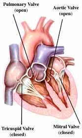
The cardiac cycle is the sequence
of events in one heartbeat.
In its simplest
form, the cardiac cycle is the simultaneous contraction of the two atria,
followed a fraction of a second latter by the simultaneous contraction of the
two ventricles.
A heartbeat
has two phases:
- Phase 1
- systole - contraction.
Occurs
when the ventricles contract, closing the AV valves and opening the SL valves
to pump blood into two major vessels leaving the heart.
- Phase 2
- diastole - relaxation.
Occurs when the ventricles relax, allowing the
back pressure of the blood to closed SL valves and opening AV valves. The
cardiac cycle also creates the heart sounds:
Each heartbeat produces two sounds, often
called lubb-dub, that can be heard with a
stethoscope.
- The first sound, the loudest and longest, is caused by the
ventricular systole (contraction) closing the AV valves.
- The second sound is caused by the closure of the aortic and
pulmonary valves (SL).
If any of the
valves do not close properly, an extra sound called a heart murmur, may be
heard.
Heart Muscle Contraction:
The heart consists of muscle cells that
contract in waves. When the first group is stimulated, they in turn
stimulate neighboring cells. Those cells stimulate more cells. This chain
reaction continues until all cells contract. The wave of activity spreads in
such a way that the atria and the ventricles contract in a steady rhythm.
The wave begins in a small bundle of
specialized heart muscle cells embedded in the right atrium called the sinoatrial node (SA). The SA-node is the
natural pacemaker of the heart. It initiates each heartbeat, without
stimulation from the nervous system, and sets the pace for the heart rate.
The impulse spreads from the pacemaker
through the cardiac muscle cells in the right and left atrium, causing both
atria to contract almost simultaneously. When the impulse initiated by the
SA-node reaches another special area of the heart known as the atrioventricular (AV) node. The AV-node is
located in the septum between the right and left ventricles. The AV-node relays
the electrical impulse to the muscle cells that make up the ventricles. The
ventricles contract almost simultaneously a fraction of a second after the
atria, completing one full heartbeat. These contractions
causes the chambers to squeeze the blood, pushing it in the proper
direction along its path.
Cardiac Output (CO) is the amount of blood pumped out of
each side of the heart (each ventricle) in 1 minute. It is the product of the
heart rate (HR) and the stroke volume (SV). Stroke volume is the volume
of blood pumped out by a ventricle with each heartbeat. In general, stroke
volume increases as the force of ventricular contraction increases.
Using the normal
resting values for heart rate (75 beats per minute) and stroke volume (70 ml -
about 2 ounces - per beat), the average adult cardiac output can be easily
figured.
CO = HR X SV
CO = (75
beats / min) ( 70 ml / beat)
CO = 5250
ml / min
Regulating
Stroke Volume:
A healthy heart pumps out about 60% of
the blood that enters it. The critical factor controlling stroke volume is how
much cardiac muscle cells are stretched just before they contract. Venous
return, the amount of blood entering the heart and distending its
ventricles, is the determining factor. Anything that increases the volume or
speed of venous return also increases stroke volume and force of contraction.
- A slow heartbeat allows more time for the ventricles to fill.
- Exercise speeds venous return because it results in increased heart
rate and force.
- The enhanced squeezing action of active skeletal muscles on veins
returning blood to the heart, the so-called muscular pump, also
plays a major role in increasing benous return.
On the other
hand, low venous return, such as might result from sever blood loss or an
extremely rapid heart rate, decreases stroke volume, causing the heart to beat
less forcefully.
Regulating Heart Rate:
For most of us, at rest our heart beats
between 60 and 80 beats per minute. Under certain conditions, that number can
increase to as many as 200 beats per minute.
- The sympathetic nervous system increases heart rate.
During times of
physical or emptional stress, the SA-node and AV-node
- and even the cardiac muscle itself - can be stimulated to increase heart
rate.
- The parasympathetic nervous system decreases it.
When demand
declines, the vagus nerves slow and steady the heart.
- Various hormones and ions can have a dramatic effect on heart rate.
Epinephrine, which mimics the effect of the
sympathetic nerves, and thyroxine both
increase heart rate.
Reduced Ca+2
in the blood depresses the heart, while a low level of
K+ causes the heart to beat feebly and without rhythm.
- A number of physical factors, including age, gender, exercise, and
body temperature, influence heart rate.
Cardiac Circulation:
Although the heart chambers are
continuously bathed with blood, the blood contained in the heart does not
nourish the myocardium. The blood supply that oxygenates and nourishes the
heart is provided by the right and left coronary arteries. The coronary
arteries branch from the base of the aorta and encircle the heart in the atrioventricular groove at the junction of
the atria and ventricles. The coronary arteries and their major branches are
compressed when the ventricles are contracting and fill when the heart is
relaxed. The myocardium is drained by several cardiac veins, which empty
into an enlarged vessel on the backside of the heart called the coronary
sinus. The coronary sinus, in turn, empties into the right atrium.
Heart-related Problems:
Pericarditis is an inflammation of the pericardium.
This can lead to a decrease in the amount of serous fluid surrounding the
heart, which in turn causes the pericardial layers to bind and stick together,
forming painful adhesions that interfere with heart movements.
Heart valves are basically simple devices, and the
heart - like any mechanical pump - can function with "leaky" valves
as long as the damage is not too great. However, severe valve deformities can
seriously hamper cardiac function.
- An incompetent valve forces the heart to pump and repump the same blood because the valve does not close
properly and blood backflows.
- In valvular stenosis,
the valve flaps become stiff, often because of repeated bacterial
infection of the endocardium (endocarditis). This forces the heart to contract more
vigorously than normal.
In each case above, the heart's workload increases, and ultimately the heart weakens and may fail. Under such conditions, the faulty valve can be replaced with a synthetic valve, or a valve taken from a pig heart.
Angian pectoris
is a crushing chest pain caused by low levels of oxygen reaching the
myocardium. While the cause of this decreased flow to the heart tissue may
vary, the pain is a warning that should never be ignored. Prolonged
angina may cause the death of ischemic (blood-starved) heart cells, forming an infarct.
The resulting myocardial infarction is commonly called a "heart
attack" or "coronary".
Fibrillation is a rapid uncoordinated shuddering of
the heart muscle. This contraction of the heart muscle is described as looking
like a "bag of worms". The cause is related to either or both of the
heart "nodes" and makes the heart totally useless as a pump. This is th major cause of death from heart
attacks in adults.
Tachycardia is a rapid heart rate (over 100 beats
per minute). Bradycardia is a heart rate that
is substantially slower than normal (less than 60 beats per minute). Neither
condition is pathological, but prolonged tachycardia may progress to
fibrillation.
Congestive heart failure (CHF) is a progressive decrease in the
efficiency of the heart. This condition reflects a weakening of the heart by
coronary artherosclerosis (clogging of the coronary
vessels with fatty buildup), persistent high blood pressure, or multiple
myocardial infarcts - leading to repair with non-contracting scar tissue.
Pulmonary congestion occurs when only the left side of the
heart fails. The right side of the heart continues to pump blood to the lungs,
but the left side is unable to send the returning blood into the systemic
circulation. As blood vessels within the lungs become swollen with blood, the
pressure causes leaking into the lung tissue, causing pulmonary edema
(lung swelling).
Peripheral congestion occurs when only the right side of the
heart fails, causing blood to back up in the systemic system. Edema is most
noticeable in the distal parts of the body - the feet, ankles, and fingers
become swollen and puffy. Failure of one side of the heart puts a greater
strain on the opposite side, and eventually the whole heart fails.